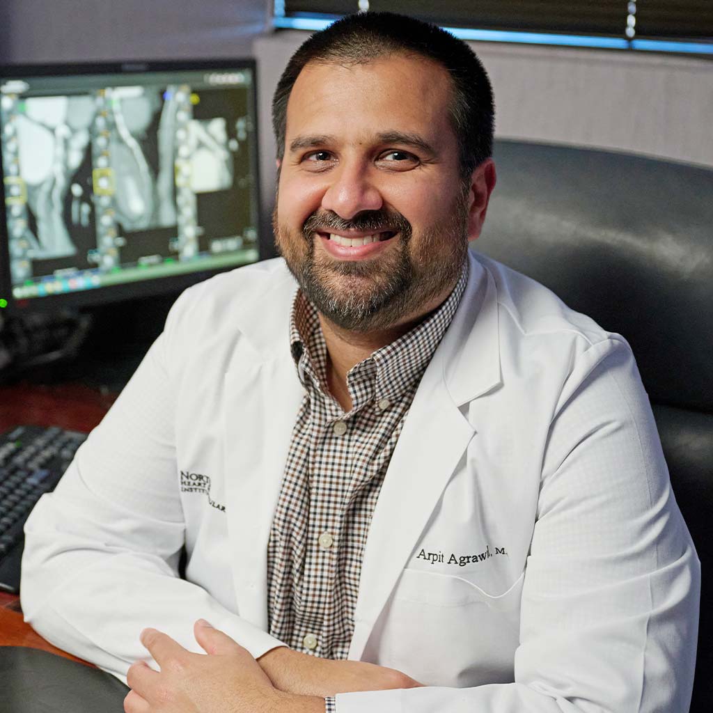
Arpit Agrawal, MD, cardiologist at Norton Heart & Vascular Institute, puts the pieces together
LOUISVILLE A good puzzle will have clues, some ambiguous, some easy to decipher. You can only solve the puzzle by putting all the pieces together, one at a time. Cardiac imaging, says Arpit Agrawal, MD, a cardiologist with a focus in cardiac imaging at Norton Heart & Vascular Institute in Louisville, is one piece of that puzzle.
“Cardiac imaging helps put the pieces of the puzzle together so we know how to treat the patient. Whether it’s with an echo or an MRI or a CT, it helps us paint the picture for that individual patient,” says Agrawal.
Agrawal was born at the downtown Norton Hospital and grew up in Louisville attending Louisville Collegiate through high school. He went to Washington University in St. Louis for his undergraduate degree and then came back to the University of Louisville for medical school. He did his internal medicine residency back at Washington University/Barnes Jewish Hospital, followed by a cardiology fellowship at Vanderbilt University Medical Center.
Agrawal was always drawn to cardiology because he was interested in engineering and cardiology was a perfect combination of the electrical activity and the pump function of the heart. “I always thought the physiology was very interesting, how the heart is a machine that works your whole life,” says Agrawal.
Agrawal joined Norton Heart & Vascular Institute in 2016. He and his wife Laila, a medical oncologist, moved back home from Nashville to be near family. “Norton Healthcare had a growth opportunity in general cardiology and in the cardiac imaging space. There was a pretty robust imaging program that has bloomed in the last several years,” he says.
A Week in the Life of a Cardiac Imaging Specialist
Agrawal has a diverse work week that includes seeing patients in the hospital or in clinic. He has dedicated time for cardiac imaging, which includes periprocedural imaging or structural imaging like transesophageal echocardiograms, which can help his interventional cardiologist colleagues during procedures such as the Watchman®, left atrial appendage occlusion, mitral clips, or transcatheter aortic valve replacements, aka TAVRs. Additionally, he spends time reading cardiac MRIs and cardiac CTs. “I like the variety I get on a day-today basis,” he says.
Heart disease can affect people of all ages, so Agrawal’s patients range from 18 years old to over 100 years old. “Heart disease affects all genders, males, females, transgender individuals. The presentations can vary. Sometimes it can be acute and dramatic presentations when they present with heart attacks, but it can also be slow and progressive such as valvular heart disease or heart failure. It can affect people no matter what stage of life they are in. Certainly, there’s a predilection to more elderly individuals, but we’re seeing younger patients with heart disease now,” he says.
Technology Advances Care
In Agrawal’s opinion, cardiac imaging is evolving and the technology is progressing quickly, particularly in coronary CT, when it’s used for evaluating patients with underlying coronary artery disease.
“There’s constant evolution in the scanner technology which reduces radiation doses while getting more precise diagnostic pictures. Recently we’ve contracted with HeartFlow® which helps us assess people’s coronaries during coronary fatigue and do some secondary analysis, which can often prevent unnecessary procedures. It’s helped our diagnostic accuracy and that’s has really changed my practice quite a bit,” says Agrawal.
Agrawal states that improved imaging has helped give specialists more confidence in stating that a blockage doesn’t need more invasive testing and is something that can be managed non-invasively. “It gives us confidence that we don’t need to proceed with an invasive procedure because the blockage is not the driver for the patient’s symptoms,” he says.
“I think cardiac imaging allows us to get data on patients that we previously were only able to get with an invasive procedure. While many cardiac invasive procedures can be done safely, anytime you are doing an invasive procedure, there’s a risk of complications. Cardiac imaging allows us to get imaging and information on patients in a much safer manner than we could previously,” says Agrawal.
Imaging and Interventional: Hand and Hand
One of the roles of the cardiac imaging specialist is working with structural interventionalists on procedures including transcatheter aortic valve replacement, left atrial appendage occlusion, and transcatheter edgeto-edge repair.
“We do the transesophageal echo during the procedure to help guide the interventionalist to put the device in the right place and gauge the success of implanting the device. And then in that same role with left atrial appendage occlusion, when we insert a Watchman device, we help guide our interventional cardiologist, making sure the device is in the right position and making sure that there’s no complications from that,” says Agrawal.
“My hope, as somebody who does cardiac imaging, is that I can help our proceduralist do procedures on the patients, guide them to make their job easier, as well as to make sure that we’re doing the right procedure for the patient. There’s strong collaboration with our cardiac surgeons, with our interventional cardiologists, and with our structural cardiologists. One of the most important things is helping patients avoid going through those processes if they don’t need it,” says Agrawal.
“The bread and butter of cardiology around the world is echocardiography, which is ultrasounds of the heart. The imaging cardiologists often read CT scans of the heart, which allow us to look at the coronary arteries in more depth. It also allows us to look at the structure of the heart in a more static manner. We also do cardiac MRI, which is a unique way of characterizing the tissue of the heart. We can see if there’s scar in the heart and what caused the scar. It can also help us get a better idea about valvular heart disease, how severe is valvular narrowing or leakiness. It also helps us look at the cardiac structures a little more in depth than what we can with echo. And then nuclear medicine is another tool that we use to help characterize tissue looking for infiltrative heart diseases such as amyloidosis or sarcoidosis, as well as another way of assessing for coronary artery disease. It kind of runs a gamut from ultrasound, CT, MRI, and nuclear medicine to help put the pieces together for the patient.”
— Arpit Agrawal, MD
The Structural Heart Team
At the Norton Heat & Vascular Institute there are typically two meetings a week that are multi-disciplinary with surgeons, imaging cardiologists, and interventional cardiologists. The structural heart meeting primarily focuses on valvular heart disease, and the team goes over all the cases under consideration for a structural heart procedure. They look at images together, including coronary angiograms, CT scans, and echocardiograms, and the multi-disciplinary team creates a comprehensive treatment plan.
The other heart team meeting is driven by the interventional cardiologists as well as general cardiologists where they go over interesting cases that some of the cardiologists have had during the week. The cardiac surgeons are also there to provide input and discuss diagnostic and surgical dilemmas.
The First Patient Consult
Solving a puzzle begins with the first piece. “I think the most important thing is listening to your patient and hearing what’s bothering them. You get so much out of hearing a patient’s story, their family history, their symptoms, how long they’ve been having symptoms. What they tell me will help guide the right diagnostic test for my patient. It also involves shared decision-making with my patient about the risks and benefits of testing and making sure that what I order aligns with how the patient and their family want to be cared for,” says Agrawal.
“Often patients and family members ask, ‘What would you do if this were your family member?’ I know that everyone’s goals are different. Some people want to live as long as they can. Some people want to just have a great quality of life for the time that they’re here. And at some point, patients’ goals change. So, when I get asked that question, it is hard to answer, unless I know exactly what the patient’s goals or their wants and needs are. What I do is get to know my patients and then recommend the test that I think that would help get them to those goals. Whether it’s an invasive test, or an imaging test, or whether it’s no test at all. Whether it’s medications, everyone’s got different goals and that’s the puzzle part about medicine: figure out what your patient’s goals are and doing what you can to align our treatment with those goals.”
“That’s one thing I enjoy doing and hope we will continue to be able to do over the course of my career,” says Agrawal.