Ankle replacement relieves pain by removing the arthritic joint surfaces. Ankle motion is maintained, leading to better walking ability and protection of adjacent joints against arthritic change over time.
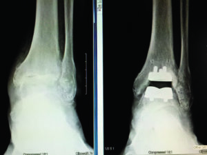
 In the top set of X-ray photos, the radiograph on the left shows the complete loss of tibiotalar joint space, which is indicative of ankle arthritis. In the ankle replacement procedure, the joint surfaces are removed with a saw and replaced by the tibial component above and the talar component below. A high-density polyethylene spacer articulates between the 2 metal components to allow ankle motion.

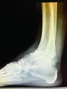
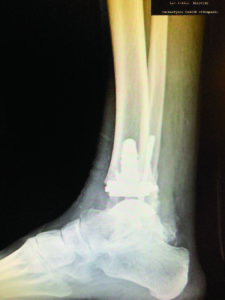
 In the bottom set of radiographic photos, the preoperative radiograph on the left shows both loss of joint space and post-traumatic recurvatum deformity. Because of previous fracture malunions of the tibia and fibula, the foot is displaced anteriorly about 2 cm. This necessitated ankle replacement as well as medial malleolar and lateral malleolar osteotomies to shift the foot back into anatomic position under the tibia. Also shown are anteroposterior prep and post-op views of the ankle showing the screws fixating the osteotomies.


